CO2 lasers and other mid-infrared light sources are proven tools for material inspection via spectroscopic techniques, recently atomic force microscopy (AFM) and scattering scanning nearfield optical microscopy (s-SNOM) have gained traction due to their complimentary setups. These techniques can provide insight to a material’s chemical composition and optical properties while providing imaging with a spatial resolution on the scale of nanometers. With the rise of metamaterials and increasingly complex nanomaterials such instruments will continue to gain prominence in both research and quality assurance.
AFM functions by measuring topographical changes in a sample due to the thermal expansion resulting from radiative absorption at various infrared (IR) wavelengths. The rapid expansion excites resonant oscillation of the AFM cantilever in contact with the surface. This rate of thermal expansion can be correlated to the chemical composition of crucial nanostructures across a diverse range of applications.
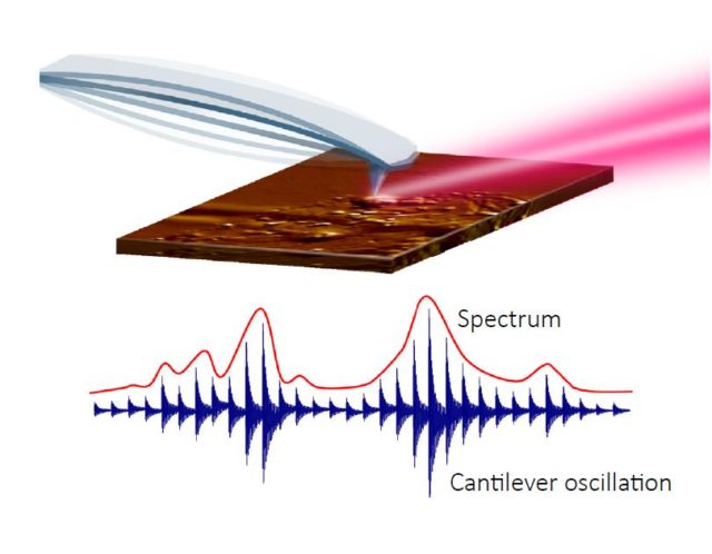
Figure 1: IR-AFM measuring wavelength dependent absorption coefficient of sample
The primary variable considered with this technique is the samples absorption spectra, IR AFM is best suited for inspection of soft materials with large thermal expansion, such as polymers and organic compounds. Providing direct measurements of infrared absorption, independent of band shifts, peak distortions and other artifacts caused by complex dependencies of tip material, geometry, and sample thickness; IR AFM correlates to conventional FTIR spectra.
In s-SNOM when incident IR light is focused onto the AFM tip apex the resultant scattered light carries the optical properties of the sample being inspected. Localized light-matter interaction under the AFM cantilever can provide spatial resolution to less than ten nanometers. Measuring both the phase of the scattered light and the optical amplitude, information regarding the complex optical constants (index of refraction n, extinction coefficient k) of both the tip and sample can be analyzed. Furthermore, the optical phase versus wavelength provides an excellent approximation to a traditional IR absorption spectrum. Due to the complexity of this signal a reference sample is often necessary for differentiating the acquired information from the source and tip. This complex and compelling technique for imaging at the nanoscale has important applications in the evaluation of advanced materials and devices. Although s-SNOM works on a wide range of materials, it has the best signal to noise ratio on harder materials with dielectric constants, high reflectivity and/or strong optical resonances.
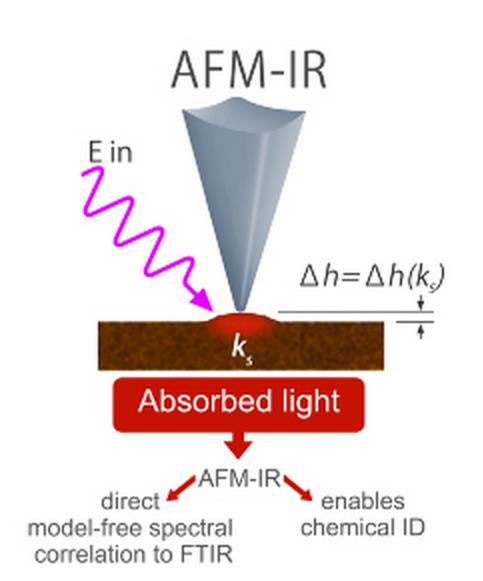
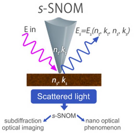
Figure 2: Comparison of the two techniques discussed.
Our L4-G and L3-LS lasers are both well suited for either of these applications. If you find yourself in a discussion and would like further clarification or technical knowledge about this or other applications, please always feel free to contact us.
Image credit to Eoghan Dillion